Current clinical guidelines for aortic valve stenosis diagnosis and intervention planning focus mainly on the pressure gradient over the valve, and on ejection fraction of the left ventricle. However, ejection fraction is merely applicable in case of global, late-stage cardiac disorders. Accordingly, left ventricular strain measured using ultrasound is a more suitable parameter. Although, time-resolved 3D ultrasound can capture the complex cardiac shape and contraction pattern, 3D+t ultrasound has a limited field-of-view, often contains near-field clutter, and has a relatively low lateral contrast and resolution.
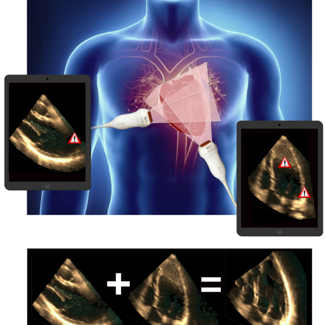
Multi-perspective ultrasound (i.e. obtaining, registering, and fusing multiple ultrasound acquisitions) increases the field-of-view, and enhances the lateral contrast and resolution. In addition, a near-field clutter reduction algorithm was created and implemented in the fusion algorithm.
Thanks to the collaboration between the TU/e, Philips, and the cardiology and radiology department of the Catharina Hospital, high-quality ultrasound images could be recorded in the Catharina Hospital that were registered using a probe-tracker designed by Philips. After fusion of the images, 3D left ventricular strains could be obtained and compared to tagged-MRI (the golden standard for strain estimation). The collaboration was especially successful due to the face-to-face contact moments, during e.g. the weekly (pre-covid) visits to Philips Research and the meetings with healthcare professionals.
klik hier